


This disease is poorly examined, although thousands of observations and subsequent treatment have been described.
The diversity and non -specificity of the pelvis varicose veins lead to severe defects from the diagnosis, which will affect the consequences in the future.
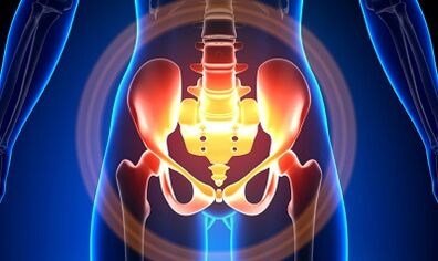
Characterization of the veins of a small pelvis
The veins of the pelvis are longer than arteries, which determine their high capacity.The reason for this is the phylogenesis of the pelvic region.The veins of the pelvis have a high adaptation capacity and are potentially prone to restructuring, which contributes to the formation of a densely intertwined network.
The velocity and direction of blood flow is regulated by the valves, which are regulated by complex humor mechanisms.Valves balance the pressure in different parts of the venous network.
When the valves cease to perform their functions, blood stagnation develops, leading to the pathology of blood vessels and the formation of varicose veins.The uniqueness of the veins of the small pelvis lies in the fact that the widespread strips of the uterus, which support the width of the ship, can also narrow it and cause pathology.
Reasons
The pathological dilatation of the veins of the pelvis may occur for the following reasons:
- Violation of blood vessels;
- Venous barrel prison;
- Compression of collateral packages with the changed position of the uterus, such as retroflexia;
- Valve ovarian failure (congenital or acquired);
- Obstructive Post -Flebitic Syndrome;
- Pathology of connective tissue;
- Arteriovenous angiodislasia;
- Prolonged seat, hard physical work;
- The varicose of the lower limbs;
- Pregnancy (3 or more) and childbirth (2 or more);
- Diseases of the female sex (chronic salpingoophorite, ovarian tumors, uterine fibroids and sexual endometriosis);
- Gluing process of pelvic organs;
- Obesity.
Classification with the degree of illness
The following degrees are different for the extended vein:
- Up to 0.5 cm, the "corkscrew" vascular stroke;
- 0.6-1 cm;
- more than 1 cm.
Possibilities of the course of illness
- The varicose of the vagina's perineum and the foyer;
- The pelvis is venous full -blooded syndrome;
Symptoms
- The most common - frequent pains in the lower abdomen, perineum after long static and dynamic surge.The pain increases in the second phase of the cycle, after hypothermia, fatigue, stress, and various diseases.
- The feeling is "not easy", pain about sex and then.
- Dysmenorrhea - menstrual disorders, including pain syndrome.
- Secretion, more than normal, gender tract bile.
- Blood stagnation leads to infertility, nonsense, and elimination of pregnancy.
- Breaking urination as a result of expanding the veins of the bladder.
Diagnosis
Diagnosis of the disease is only successful based on complaints in only 10 % of cases.
The palpation of the inner walls of the pelvis allows you to feel the nodes of elongated seals and veins.Examining the mirrors, you can see cyanosis of the vaginal mucosa.
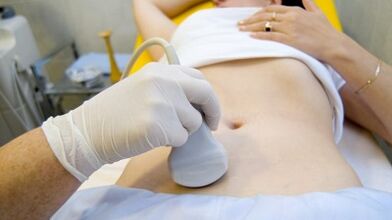
The choice procedure is an ultrasonic study that has a colorful doppler map that allows not only the varicose veins of the ovarian veins, but also to identify venous thrombosis, post -romboflitetic obstruction.Ultrasound, "worms", without reflecting the signal, localized on the side surface of the uterus.
The effect of the Doppler study is blue and red "coloring", venous and arterial blood that corresponds.
The device for ultrasonic examination recognizes blood movement from the sensor with a special program and calculates the blood flow rate and the type of blood in the other direction.
But the exact determination of the vein or the artery remains behind the doctor.The doppler method works almost always, and our body dictates the exceptions of the rules because the blood from the heart is not always arterial and vice versa.
Thus, an ultrasonic diagnostic physician sees arterial or venous blood vessels, size, blood flow rate and many indicators that an ordinary person does not need, but plays an important role in the diagnosis.To do this, use transbdominal and transvaginal sensors.
In 5.7% of the cases, the disease is recognized in an accident during screening.Generally, the vena ovarian diameter is 0.4 cm.
CT and MRI have high accuracy.Using these methods, the accumulation of varicos can be found in the leagues around the uterus, ovary and organs.Concomitant pathology can be determined.
A very reliable method is a fleet study.
The contrast is performed at the height of the valsalva pattern against blood flow.This allows the valve to fail.
Retronenoscopy on the left side is used by kidney fleboraogography, super -sequenic fleebovarioscopy and fleeboovariography on both sides.These methods allow them to determine the hemodnam and anatomical changes of the veins of the kidneys and the location of falling into the gonadny blood vessels.
The super -publisher's fleeravarioscopy is performed by catheterization of the gonad veins through a controversial femur or sub -calvian vein, later introduced by the contrast.
Most of the blood from a hornless plexus is thrown away by the ovarian vein.But under high blood pressure conditions, non -cornuted uterus occurs into the inner iliac veins.The plexus of the veins through which the outflow can occur includes the sacral and plexus of the bladder.
In the left -side Flebo cigar, 3 stages of venous stagnation are distinguished in the pulp cluster plexus of the left side:
- There is no outflow from the plexus of the left -hand ovary, otherwise it will be made along a further short way.
- There is a further long way.
- Two additional outflow paths or one additional and additional care.
In sections 2 and 3, the right ovarian cluster plexus is formed.
Laparoscopy is used for differential diagnosis.The pathologically winding veins are in the ovary towards the round and wide ribbons.They look like a large cyanotic conglomerate with a thin and tense wall.
The complexity of the diagnosis lies in the fact that the disease is often hidden behind signs of the inflammatory process, distinguished by endometriosis, prolapse of the internal organs, postoperative neuropathies and many extragenital diseases mask.
Treatment
The main purpose of the treatment is to remove the veins' reflux.Conservative treatment is used in the initial stages of the disease.In the late stages of the disease, the choice of treatment is surgery.
Conservative treatment

This consists of normalizing the sound of veins, improving hemodynamics and trophic processes.
Symptomatic treatment to eliminate each symptom.Non -steroid anti -inflammatory pain, bleeding - hemostatic therapy.
The main drugs of conservative treatment are venotonic drugs and antiplatures.
Flebotonics - Improve the sound of the vascular wall and improve bloodstream.With this disease, it is better to consult a gynecologist about certain drugs.
Physiotherapy exercises are an important method.
Surgery
- Revival varicos.
- Gonado-Cavalry Shunting.
- Sclerosis for laparoscopy.
- Occrames of the ovarian veins by X-ray-endovascular methods.
Folk medicines
Because the most important thing in the occurrence of the disease is the weakness of the valve device, this pathology is also used for all folk drugs used to expand the veins of the lower limbs.
The most commonly used hazelnuts, hops, nettle, horse chestnut, dandelion root, tea mushrooms, willow, oak, St. John's Wort, series, pollen powder and many other plants.
Effective: Bag baths, chestnuts, willows, chamomile, pharmacy, drying, treatment of St. John's beer.
Prevention
- The first thing when complaints, predictors or illnesses are listed above - contact a gynecologist.
- Normalize the working system normalization and relaxation, try not to stay upright for a long time.
- Do exercises to prevent “pedal”, “stand-up stack”, “leg feet”
- Stick to the diet: eat foods with high content with vitamin E, R, C, try to eat white meat, less fat, replace fruits, vegetables, cereals.
- Drink sufficient amounts of liquid, but at least 1.5 liters per day.
- Get rid of overweight, bad habits.
- Consult your doctor to wear the compression canvas, which improves blood outflow from the lower limbs, thus less stagnation in the pelvis.
- Avoid baths, sauna, steam bath, hot bath.
In order not to be ill with such a serious illness, the preventive recommendations listed above must be followed.Treat your health in life the most valuable.
For the smallest suspicious symptoms that you should not get rid of for several days, consult a doctor.He must provide highly skilled help and save him from suffering.